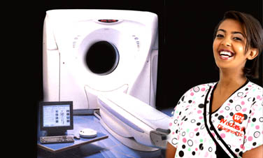
CT SCAN
Computed Tomography (CT) is an advanced technology producing images of the body much like the slicing of a loaf of bread. It is a highly sensitive method to accurately view the internal anatomy and detect extremely small lesions. Utilizing high speed computers, the CT machine obtains 360 degrees of X-ray information. This is processed into single slice images for display on a monitor and can be reproduced on film or high quality photographic paper.
Our multi-slice ELSCINT light speed machine with helical scanning capability significantly shortens examination time and reduces the volume of intravenous non-ionic contrast material administered while providing exceptional resolution. During a patient's single breath hold, helical scanning produces multiple contiguous slices so that extremely small lesions are not obscured. These features enhance diagnostic sensitivity, early detection, and improve consistency of follow-up pathology while ensuring patient comfort.
During the scan, you will be asked to rest motionless on a padded table for 5 to 15 minutes depending on the area to be scanned. The table moves every few seconds as the images are obtained. You will hear faint humming and clicking sounds. After the images are taken, a radiologist will review the images to make sure all of the area has been covered. Because of breathing or motion inside the body, additional images are sometimes needed. Additional images do not mean there is a problem.
Depending on the part of the body being scanned, different contrast materials are used. Contrast is often administered through the vein (intravenous). The contrast we use is Iopamiro or Ultravist 370mg or 300mg depending on the part of the body being scanned. Although reactions are quite rare, it is not unusual to get a flush feeling during the exam or a metallic taste in the mouth. This typically lasts for less than a minute.
If you have an iodine allergy or have had a reaction to contrast in the past, you should notify the office when you are making the appointment, and also at the time of the scan, so that we can take additional precautions to avoid a problem. If you are taking a diabetic medication called Glucophage, please alert us at the time of your appointment, as this may react with the IV iodine and result in kidney problems.
Oral contrast is usually given for CT scans that include the abdomen and pelvis. The oral contrast is swallowed and courses throughout the gastrointestinal tract. The intestines then appear white on the CT images.
Two types of substances serve as oral CT contrast. Barium sulfate is similar in consistency to a milkshake. Gastrograffin or Urograpin is a water-based drink containing iodine and is generally mixed with pure water. Patients usually need to drink about 32 oz. of either contrast to adequately fill the stomach and intestines. Minor side effects such as constipation or diarrhea may occur.
For CT examinations that include the pelvis, you will be asked to arrive three hours before the actual scan time to drink the contrast. For a CT scan of the abdomen you will be scheduled to arrive two hour before scanning. This is necessary to allow adequate filling of the intestines, which helps in the proper interpretation of the study. You will be seated in our waiting room during this period. Reading material will be available, however, feel free to bring your own.
Screening examinations have become important parts of healthcare, used to detect disease before they become clinically problematic. Currently, screening chest CT for the early detection of lung cancer and heart disease are simple non-invasive exams that we perform in our diagnostic center.
CT Preparation Instructions
Computed Tomography Scan of Body Parts |
Preparation |
CT Head/Neck or Chest: |
Nothing to eat or drink four hours prior to examination |
Abdomen and/or Pelvis |
Clear liquid diet for 4 hours prior to exam. Liquids include clear juice such as apple, cranberry and grape, clear soups, Jell-O, coffee, or tea. No milk products or carbonated beverages. |
Spine/Bone or Joint |
No preparation necessary. |
POST PROCEDURE INSTRUCTIONS:
BARIUM STUDY: Following a barium study, it is recommended that you take 3 tea spoons of milk of magnesia the evening of your exam or at bedtime. It is also recommended that you increase your daily fluid intake.
If you do not have a bowel movement within 24 hours, repeat the dose and please contact our office.
CONTRAST CT: Following your CT scan, it is recommended that you increase your fluid intake by 4 glasses on the day of your examination. You may have watery bowel movements for 24 hours.
CT Myelograms
A Computerized Tomography (CT) scan can help diagnose different spinal conditions including disc herniation, spinal stenosis, tumor, and vertebral fracture. CT provides a radiographic image of a single body plane. It is particularly good at imaging hard tissue, such as bones. The equipment is shaped like a donut or ring, with a movable table that slides in and out of the ring. The scanning system includes a computer that creates cross-sections or slices of a specific part of the spine. Obtaining similar images is not possible with traditional X-rays.
A Myelogram (also known as myelography) is a diagnostic tool that uses radiographic contrast media (dye) that is injected into the spinal canal’s fluid. After the dye is injected, the contrast dye serves to illuminate the spinal canal, cord, and nerve roots during imaging.
Thus, when a CT scan and myelography are combined, images are produced that clearly show both the bony structures of the spine and the nerve structures. These images are invaluable to physicians as they diagnose a patient’s spinal problem condition.
Testimonial

“I never thought that my child would get a chance to be diagnosed and treated. I saw no hope. I’m really glad to find such a service in your organization. When my child’s health is restored, I’ll be able to do much more.”
His mom: Bedria Reshid
Patient: Abdela Nasir
Get in Touch
Address: P.O.Box. 5735 Addis Ababa Ethiopia
Location: Churchill Road in front of Lion Pharmacy
Telephone: +251-111-574343
FAX: +251-111-553599
Mob: +251-911-240182
E-mail: info@wudassiediagnostic.com
Website: www.wudassiediagnostic.com